|
??? |

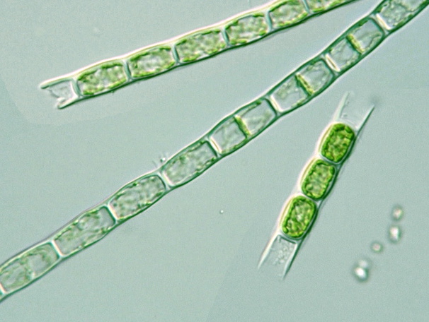
Microspora sp.
52-80 x 25-30 μm
Iruma river
Kawagoe
Saitama, 2004 |

Microspora sp.
27-54 x 30 μm
Tama-gawa g.p.
Fuchu
Tokyo, 2004 |

Microspora sp.
46 x 30 μm
Higusa-numa
Hikari
Chiba, 2006
 |

Microspora sp.
43-65 x 25 μm
Toneri Park 2
Adachi-ku
Tokyo, 2006
|

Microspora sp.
μm
Water reservoir
Ohira
Tochigi, 2008
|

Microspora sp.
40-80 x 25 μm
Hamayu-no-mori
Izumo
Shimane, 2006
|

Microspora sp.
24-48 x 25 μm
Yanase river
Tokorozawa
Saitama, 2004 |

Microspora sp.
17-35 x 20 μm
Kata-numa
Narugo onsen
Miyagi, 2006
|

Microspora sp.
15-20 x 17 μm
Chigo-ike 4
Shiga highland
Nagano, 2006
|
|
M. pachyderma: Cell body 6.6-13 μm in diam., length 1.3-3 times longer than witdh;
H structure conspicuous, cell wall 1.5-2.5 μm in thickness |

M. pachyderma
17 samples
8-28 x 11-14 μm
2005-2008 |

M. pachyderma
17 samples
8-28 x 11-14 μm
2005-2008 |

M. pachyderma
17 samples
8-28 x 11-14 μm
2005-2008 |

M. pachyderma
17 samples
8-28 x 11-14 μm
2005-2008 |
|
M. willeana: Cell body 10-12 μm in diam., length 13-18 μm; |

M. willeana
8 samples
9-20 x 9-14 μm
2004-2007 |

M. willeana
8 samples
9-20 x 9-14 μm
2004-2007 |

M. willeana
8 samples
9-20 x 9-14 μm
2004-2007 |

M. willeana
8 samples
9-20 x 9-14 μm
2004-2007 |
|
M. tumidula: Cell body 8-9 μm in diam., length nearly equal to its width; |

M. tumidula ?
μm
Komado marsh
Showa and Tajima
Fukushima, 2004 |

M. tumidula ?
6-7 x 8 μm
Komado marsh
Showa and Tajima
Fukushima, 2004 |

M. tumidula ?
6-11 x 8 μm
Kisuge konuma
Shimogo
Fukushima, 2005 |

M. tumidula ?
5-10 x 9 μm
Onnenai
Kushiro marsh
Hokkaido, 2005 |

M. tumidula ?
6.5-13 x 8.5 μm
Midoriga-ike
Togakushi highland
Nagano, 2006
|

M. tumidula ?
x 8-9 μm
Kuroyachi marsh
Hachimantai
Iwate, 2007
|

Microspora sp. ?
8-18 x 9 μm
near Iroha-numa
Zao mountain
Yamagata, 2006
|

Microspora sp.
13-16 x 8 μm
Numa-no-hara m.
Madarao highland
Niigata, 2005 |

Microspora sp.
16-20 x 9 μm
Kusatsu Onsen b.t.
Kusatsu
Gunma, 2005 |

Microspora sp.
8-15 x 9 μm
Karakemi marsh
Yasaka
Nagano, 2005 |

Microspora sp.
12-17 x 5-9 μm
Karakemi marsh
Yasaka
Nagano, 2006
|

Microspora sp.
6-14 x 8.5 μm
Iyari marsh
Omachi
Nagano, 2006
|
|
M. stagnorum: Cell body 5-7 μm in diam., length 1-3 times longer than witdh |

M. stagnorum ?
15-19 x 7 μm
Onnenai
Kushiro marsh
Hokkaido, 2005 |

M. stagnorum ?
4.5-9 x 7 μm
Toyonodai park
Otone
Saitama, 2006
|

M. stagnorum ?
5-9 x 5.5 μm
Nidanuma
Tsuchiyu-onsen
Fukushima, 2005 |
|
??: Cell body narrower than 5 μm in diam. |

Microspora sp. ?
4-9 x 4 μm
Onnenai
Kushiro marsh
Hokkaido, 2005 |
|
Microspora or Geminella or ? |

Microspora sp.
or Geminella ?
4-8 x 7 μm
Lake Nojiri
Shinano
Nagano, 2004 |

Microspora sp.
or Geminella ?
6-7 x 8 μm
Riverside green l.
Sawara
Chiba, 2004 |

Microspora sp.
or Geminella ?
6-7 x 8 μm
Numa-no-hara m.
Madarao highland
Niigata, 2005 |

Microspora sp.
or Geminella ?
6-12 x 7 μm
Tanohara Natural p.
Ontake highland
Nagano, 2005 |

Microspora sp. or
Klebsormidium ?
9-18 x 7 μm
Karakemi marsh
Yasaka
Nagano, 2006
|