 Genus: Similar to Chlamydomonas, but pyrenoids absent.
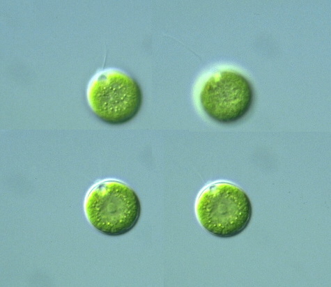
Species: Cell body spherical, 12-25 μm in diam. (young cell slightly ellipsoidal); papilla broad conical in shape; nucleus centrally located; a single chloroplast cup-shaped; stigma large, elliptical or circular in shape, located at anterior half of the cell; 2 contractile vacuoles at the base of flagella (Süßwasserflora von Mitteleuropa 9, Chlorophyta I, 1983). |


Chloromonas maculata Korschikoff (Syn: Chamydomonas korschkoffii Pascher): Cell body spherical, 15-26 μm in diam.; 2 flagella approximately equal to cell-body length; papilla small, flat in shape; many chloroplast small plate-like, without pyrenoids; stigma elliptical; 2 contractile vacuoles at the base of flagella; nucleus located at the center of the cell body (Photomicrographs of the Freshwater Algae, vol. 17, 1996). Cell body 15-22 μm in diam.; many plate-like chloroplasts circular or polygonal in shape (Süßwasserflora von Mitteleuropa 9, Chlorophyta I, 1983).
Chloromonas ulla (Skuja) Gerloff et Ettl: Cell body spherical, 12-25 μm in diam. (young cell slightly ellipsoidal); papillae half-circular in shape; nucleus centrally located; a single chloroplast cup-shaped; stigma large, elliptical or circular in shape; 2 contractile vacuoles at the base of flagella (Süßwasserflora von Mitteleuropa 9, Chlorophyta I, 1983).