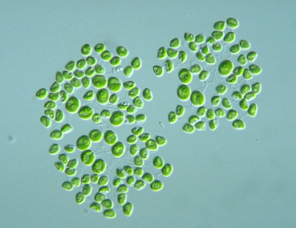
| Cell body cup-shaped | |||

D. reniforme cell 7 x 4 μm Japan, 1998 |

D. reniforme cell 9 x 6.5 μm Uruguay, 1999 |

D. reniforme ? cell 10 x 6 μm Japan, 2000 |

D. reniforme cell 7 x 5 μm Kawagoe Saitama, 2001 |

D. reniforme cell 9 x 7 μm Mizumoto park Tokyo, 2001 |

D. reniforme cell 8 x 6 μm Mizumoto park Tokyo, 2001 |

D. reniforme cell 7 x 5 μm Kasuga Fukuoka, 2001 |

D. reniforme cell 9 x 8 μm Nasu Tochigi, 2001 |

D. reniforme ? cell μm Sayama Saitama, 2001 |

D. reniforme stock Ssa-3, 5, 9 cell 7 x 6 μm Sayama Saitama, 2001 |

D. reniforme cell 8 x 7 μm Otto-Numa Tsuchiura Ibaraki, 2002 |

D. reniforme cell 8 x 7 μm Heisei-no-mori Park Kawashima or Kawajima Saitama, 2002 |

D. reniforme cell 7 x 4 μm Otto-Numa Tsuchiura Ibaraki, 2002 |

D. reniforme cell μm Iruma River side Tsurugashima Saitama, 2003 |

D. reniforme cell μm O-ike park Yabuki Fukushima, 2003 |

D. reniforme cell μm Higusa-numa Hikari Chiba, 2003 |

D. reniforme cell μm Aobano-mori p. Chiba Chiba, 2003 |

D. reniforme ? μm φ Kijioka Castle Ruin Kodama Saitama, 2003 |

D. reniforme ? 3-5 x 5-6 μm Hasu-ike Shiga highland Nagano, 2005 |

D. reniforme ? 6 x 7 μm Tane-ike Togakushi highland Nagano, 2005 |

D. reniforme cell 7 x 5 μm Tane-ike Togakushi highland Nagano, 2005 |
|||
