 Genus: Striated sutural regions dorsal and to rear of cytoproct; body tapers to rear from 3 sides
(Illustrated Guide, 1985).
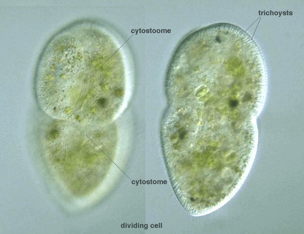
Broadly rounded anterior end and bluntly pointed narrow posterior end; sausage-form macronucleus;
a micronucleus; contractile vacuole middle of body with collecting canals; fresh water (Kudo, 1966).
Genus: Striated sutural regions dorsal and to rear of cytoproct; body tapers to rear from 3 sides
(Illustrated Guide, 1985).
Broadly rounded anterior end and bluntly pointed narrow posterior end; sausage-form macronucleus;
a micronucleus; contractile vacuole middle of body with collecting canals; fresh water (Kudo, 1966).
Buccal structure is well-stained by azure C (Y. Tsukii, 2000). Species: Cell body 140-155 μm; long (Kahl, 1930). |


D. bütschlii, Lauterborn:
135-155 μm long; with or without zoochlorella;
fresh water (Kudo, 1966).
D. colpidioides von Gelei: Reniform, twisted;
100-160 μm long (Kudo, 1966).