
Lobosea:
Gymnamoebia:
Amoebida: Thecina: Striamoebidae (or Thecamoebidae)
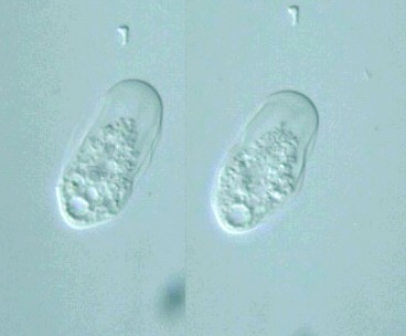
Striamoeba quadrilineata
or Thecamoeba quadrilinaeta
(Carter, 1856) Lepsi, 1960
 Family & Genus: Under 100 μm long;
oblong to ovate; with several, longitudinal, parallel ridges; uninucleate .
Species: Locomotive, 40-50 μm long,
30-40 μm wide; pseudopods none; uroid none;
ectoplasm, pale, as anterolateral margin; 3-4 dorsal ridges; endoplasm finely granular;
nucleus spherical 10 μm, endosome ring between
2 polar caps; contractile vacuole none ? (Illustrated Guide, 1985).
|
Similar species ->> Thecamoeba striata
Striamoeba quadrilinaeta,
cell body 42 μm long, 21 μm wide,
nucleus compact- or slightly vesicular-type,
x 640, Shinobazu-ike (pond), Ueno, Taito-ku, Tokyo, Japan, March 2001 by Y. Tsukii
 31 μm
31 μm
 63 μm
63 μm
 94 μm; x 640


S. munda Penard, 1890: Locomotive, 40-50 μm long,
30-40 μm wide; pseudopods none; uroid none;
ectoplasm, pale, as anterolateral margin; 3-4 dorsal ridges; endoplasm finely granular;
nucleus spherical 10 μm, endosome ring between
2 polar caps; contractile vacuole none ? (Illustrated Guide, 1985).
S. similis (Schaeffer, 1926): 30-80 μm long;
broadly ovate, with few dorsal ridges; nuclear endosomal granules scattered under nuclear
membrane (How to know the protozoa, 1979).
S. quadrilineata: 35-38 μm long;
elliptical; 2-5, usually 4 dorsal ridges; nucleus with single, smooth endosome
(How to know the protozoa, 1979).
S. striata (Penard, 1890): Locomotive, 30-50 μm long, 20-30 μm wide,
pseudopods rare; uroid none; ectoplasm pale; 3-5 dorsal ridges and clear, antero-lateral margin;
endoplasm finely granular; nucleus spherical, 5-6 μm, endosome as a band or 2-3 pieces;
contractile vacuole 8-12 μm (Illustrated Guide, 1985).
Ovate; 30-80 μm long; 3-4 dorsal ridges;
nucleus with 2 or 3 ribbon-like endosomes (How to know the protozoa, 1979).
S. corrugata : 35-80 μm long;
usually 6 ridges; slightly tapered front and rear; nucleus with plastic central endosome
(How to know the protozoa, 1979).
Please click on images for viewing enlarged.
Copyright
Protist Information Server
 Family & Genus: Under 100 μm long;
oblong to ovate; with several, longitudinal, parallel ridges; uninucleate .

