 Genus: Test with 2 pseudostomes; with thin, organic test (Illustrated Guide, 1985).
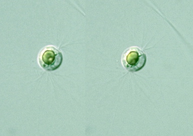
Species: Colonial or solitary; cell body spherical (8-20 μm in diam.), covered with a delicate hyaline membrane; two pseudostomes situated at or near two opposite poles; cytoplasm colorless, transparent, finely granular, filling the envelope (test) except near the two pseudostomes; a single nucleus with a nucleolus; one large, or two or three small, colored, oil-like globules present; pseudopodia extremely attenuate, radiating, straight or dichotomously branced, emanating each pseudostome; reproduction by fission or tetrad division (Cash, J. (1915) the British Freshwater Rhizopoda and Heliozoa, III, p. 145.) |


Diplophrys archeri Barker:
Colonial or solitary; cell body spherical (8-20 μm in diam.), covered with a delicate hyaline membrane;
two pseudostomes situated at or near two opposite poles; cytoplasm colorless, transparent, finely granular, filling the
envelope (test) except near the two pseudostomes; a single nucleus with a nucleolus;
one large, or two or three small, colored, oil-like globules present;
pseudopodia extremely attenuate, radiating, straight or dichotomously branced, emanating each pseudostome;
reproduction by fission or tetrad division
Young cells frequently aggregated into colonies which form circular masses about 30-60 μm in diam. or more;
from the perphery of which slender pseudopodia radiate;
Cells in a colony about 4 μm in diam., colorless, and transparent except for one dark spot in each
(Cash, J. (1915) the British Freshwater Rhizopoda and Heliozoa, III, p. 145.)
Barker (1868) Qrt. Jrn. Micr. Sci. VIII, p. 123; Barker (1869) Proc. Dubl. Micr. Club, I, 3, p. 178.