 Species:
80-170 μm long,
25-45 μm wide;
posterior bluntly rounded; flagellum about the body length; pellicle striated;
chloroplasts elongated, parallel to the striae;
haematochrome granules scattered in sun light and gathered in central area in darkness
(Kudo, 1966).
Body broadly fusiform;
90-120 μm long,
25-35 μm wide;
anterior rounded, posterior tapered with a short process;
chloroplasts tasselized and forming chromatophore bands, 12-16 in number;
flagellum = body length
(Photomicrographs of the Freshwater Algae, 1996).
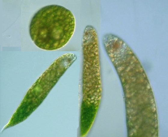
Species:
80-170 μm long,
25-45 μm wide;
posterior bluntly rounded; flagellum about the body length; pellicle striated;
chloroplasts elongated, parallel to the striae;
haematochrome granules scattered in sun light and gathered in central area in darkness
(Kudo, 1966).
Body broadly fusiform;
90-120 μm long,
25-35 μm wide;
anterior rounded, posterior tapered with a short process;
chloroplasts tasselized and forming chromatophore bands, 12-16 in number;
flagellum = body length
(Photomicrographs of the Freshwater Algae, 1996).
|


Euglena rubra Hardy: 70-170 μm long, 25-36 μm wide; cell body cylindrical; rounded anteriorly & tapered posteriorly; spiral stiation; nucleus posterior; flagellum longer than body; stigma about 7 μm in diam., many fusiform chloroplasts aligned with the striae; numerous haematochrome granules; paramylum bodies ovoid (Kudo, 1966).
* Euglena sanguinea Ehrenberg, 1830 (Syn. E. haematodes (Ehrenberg, 1830) Lemmermann, 1913): 80-170 μm long, 25-45 μm wide; posterior bluntly rounded; flagellum about the body length; pellicle striated; chloroplasts elongated, parallel to the striae; haematochrome granules scattered in sun light and gathered in central area in darkness (Kudo, 1966). Body broadly fusiform; 90-120 μm long, 25-35 μm wide; anterior rounded, posterior tapered with a short process; chloroplasts tasselized and forming chromatophore bands, 12-16 in number; flagellum = body length (Photomicrographs of the Freshwater Algae, 1996).