 Genus: Buccal cavity and internal atrium large; a well-developed cytopharyngeal basket present;
the atrial depression is in two parts, the outer shallow region and an inner deep region leading to
the cytostome (Carey, 1992). Like Nassula, but
cyrtos has smaller opening (How to know the protozoa, 1979).
Genus: Buccal cavity and internal atrium large; a well-developed cytopharyngeal basket present;
the atrial depression is in two parts, the outer shallow region and an inner deep region leading to
the cytostome (Carey, 1992). Like Nassula, but
cyrtos has smaller opening (How to know the protozoa, 1979).
Species: |


Images of collecting locality:
No.1, Kannon-numa Forest Park (pond), Shimogo town, Fukushima Pref., Japan, November 15, 2008, by Y. Tsukii


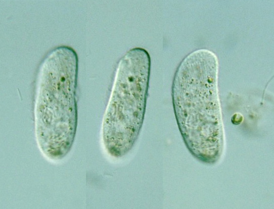
P. microstoma (Claparède and Lachmann): Pellicle roughened by a criss-cross of longitudinal and circular furrows; macronucleus elongate oval, posterior; contractile vacuole middle & right-dorsal; about 80-95 μm long, salt water (Kudo, 1966).
P. brunnea (Fabre-Domergue, 1885) Fauré-Fremiet, 1963: 250-300 μm long; flexible and elastic but non-contractile; a single contractile vacuole middle; macronucleus long and vermiform (Carey, 1992).