 Genus: Postoral kineties usually to left of oral poykinetids (Illustrated Guide, 1985).
Left edge is more curved than right edge; cytopharynx with numerous strong fibrils; ectoplasm with
numerous fusiform trichocysts; macronucleus oval; one to several micronulei (Kudo, 1966).
Genus: Postoral kineties usually to left of oral poykinetids (Illustrated Guide, 1985).
Left edge is more curved than right edge; cytopharynx with numerous strong fibrils; ectoplasm with
numerous fusiform trichocysts; macronucleus oval; one to several micronulei (Kudo, 1966).
Species: 150-600 μm long; ventrally flattened; dark colored; buccal cavity close to anterior end (How to know the protozoa, 1979). Elongate 150-600 μm long; anterior is not wider than posterior; cytostome small with well defined pre- and postoral suture; a large macronucleus with three micronuclei; single contractile vacuole with 12 radiating canals; many trichocysts (Carey, 1992). Cell body 150-600 μm long (Kahl, 1930). |


Frontonia leucas, stock Nnm-1,
cell body 280 μm long, 120 μm wide,
a single macronucleus ellipsoidal, 60 μm long, 30 μm wide,
trichocysts 8 μm long, 1 μm wide, cytostome 40 μm long,
x 100, x 200, x 400, x 640, Matsumoto, Nagano Pref., Japan, 2001 by Y. Tsukii
![]() 50 μm
50 μm
![]() 100 μm
100 μm
![]() 150 μm; x 400
150 μm; x 400


![]() 31 μm
31 μm
![]() 63 μm
63 μm
![]() 94 μm; x 640
94 μm; x 640


Frontonia leucas, stock Nnm-1,
cell body 320 μm long, 100 μm wide,
x 100, x 200, x 400, x 640, Matsumoto, Nagano Pref., 2001 by Y. Tsukii
![]() 50 μm
50 μm
![]() 100 μm
100 μm
![]() 150 μm; x 400
150 μm; x 400


Cytostome, x 640
![]() 31 μm
31 μm
![]() 63 μm
63 μm
![]() 94 μm; x 640
94 μm; x 640


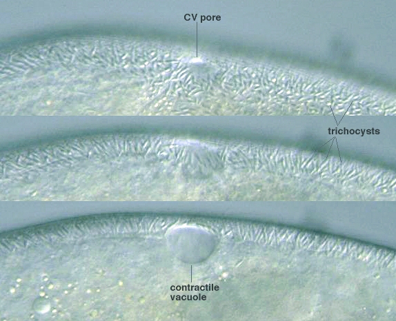
Contractile vacouole pore and radial canals
![]() 31 μm
31 μm
![]() 63 μm
63 μm
![]() 94 μm; x 640
94 μm; x 640


Contractile vacouole pore,
cell body 280 μm long, 110 μm wide,
x 100, x 200, x 400, x 640


Macronucleus 63 μm long, 27 μm wide, with micronuclei, x 640

