
Oligohymenophorea
(formerly Nassophorea): Peniculida: Frontoniina: Frontoniidae
Frontonia depressa
(Stokes, 1886)
Frontonia parvula Penard, 1922
 Genus: Postoral kineties usually to left of oral poykinetids (Illustrated Guide, 1985).
Left edge is more curved than right edge; cytopharynx with numerous strong fibrils; ectoplasm with
numerous fusiform trichocysts; macronucleus oval; one to several micronulei (Kudo, 1966).
Species:
Cell body 60-80 μm long; macronucleus short sausage-shaped; a single micronucleus; a single contractile vacuole
(Kahl, 1930).
|
Similar Genus --> Disematostoma
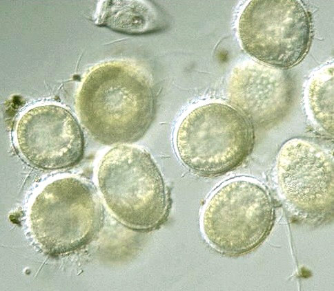
Frontonia depressa,
cysts 32 μm in diam.,
x 400, x 640, Japan, December 1998, by Y. Tsukii
 50 μm
50 μm
 100 μm
 150 μm; x 400 :
 31 μm
31 μm
 63 μm
63 μm
 94 μm; x 640


* F. depressa (Stokes, 1886) (F. parvula Penard, 1922):
Cell body 60-80 μm long; macronucleus short sausage-shaped; a single micronucleus; a single contractile vacuole
(Kahl, 1930).
F. acuminata Ehrenberg, 1833
(F. acuminata var. angustata Kahl, 1931; F. arenaria Kahl, 1933):
Ovoid, 60-100 μm long; cytostome ovoid;
a single macronucleus and single micronucleus anterior; a single contractile vacuole central;
numerous large trichocysts (Carey, 1992).
F. macrostoma Dragesco, 1960:
Ovoid, small 125-230 μm long;
very large oral aperture, well defined suture; a large macronucleus with single micronucleus;
a contractile vacuole posterior; many large trichocysts; pellicle with interkinetal striation
(Carey, 1992)
Frontonia elliptica Beardsley, 1902:
Cell body 150-200 μm long; macronucleus ovoid, with a single micronucleus large, ovoid in shape;
two contractile vacuoles present (Kahl, 1930).
Frontonia fusca (Quennerstedt 1869):
Cell body 150-200 μm long; macronucleus ovoid, with a small micronucleu;
a single contractile vacuole located at posterior half of the cell body
(Kahl, 1930).
Please click on images for viewing enlarged.
Copyright
Protist Information Server
 Genus: Postoral kineties usually to left of oral poykinetids (Illustrated Guide, 1985).
Left edge is more curved than right edge; cytopharynx with numerous strong fibrils; ectoplasm with
numerous fusiform trichocysts; macronucleus oval; one to several micronulei (Kudo, 1966).


