 Genus: Striated sutural regions dorsal and to rear of cytoproct; body tapers to rear from 3 sides
(Illustrated Guide, 1985).
Broadly rounded anterior end and bluntly pointed narrow posterior end; sausage-form macronucleus;
a micronucleus; contractile vacuole middle of body with collecting canals; fresh water (Kudo, 1966).
Genus: Striated sutural regions dorsal and to rear of cytoproct; body tapers to rear from 3 sides
(Illustrated Guide, 1985).
Broadly rounded anterior end and bluntly pointed narrow posterior end; sausage-form macronucleus;
a micronucleus; contractile vacuole middle of body with collecting canals; fresh water (Kudo, 1966).
Buccal structure is well-stained by azure C (Y. Tsukii, 2000). Species: Cell body 140-155 μm; long (Kahl, 1930). |


Disematostoma small variety ?, tapered at rear,
cell body 150 μm long, 98 μm wide,
x 100, x 200, x 400, August 7-13, 1999, Vermont-Maine U.S.A., by Y. Tsukii
![]() 50 μm
50 μm
![]() 100 μm
100 μm
![]() 150 μm; x 400
150 μm; x 400


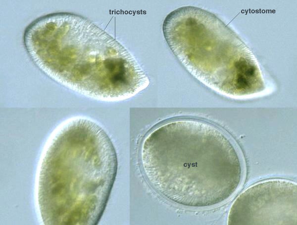
Disematostoma bütschlii, cysts,
cell body 103 μm long, 86 μm wide,
x 100, x 200, x 400, x 640, August 7-13, 1999, Vermont-Maine U.S.A., by Y. Tsukii
![]() 50 μm
50 μm
![]() 100 μm
100 μm
![]() 150 μm; x 400 :
150 μm; x 400 :
![]() 31 μm
31 μm
![]() 63 μm
63 μm
![]() 94 μm; x 640
94 μm; x 640


D. bütschlii, Lauterborn:
135-155 μm long; with or without zoochlorella;
fresh water (Kudo, 1966).
D. colpidioides von Gelei: Reniform, twisted;
100-160 μm long (Kudo, 1966).