 Species:
Colonies of 128 cells, of which 48-64 cells (anterior 1/3~1/2 of the cells in a colony) small,
8-15 μm in diam., others large, 30-35 μm in diam.;
colonies spherical in shape, 150-200 μm in diam.; a cup-shaped chloroplast with a pyrenoid;
(Illustrations of The Japanese Fresh-water Algae, 1977).
Species:
Colonies of 128 cells, of which 48-64 cells (anterior 1/3~1/2 of the cells in a colony) small,
8-15 μm in diam., others large, 30-35 μm in diam.;
colonies spherical in shape, 150-200 μm in diam.; a cup-shaped chloroplast with a pyrenoid;
(Illustrations of The Japanese Fresh-water Algae, 1977).
|


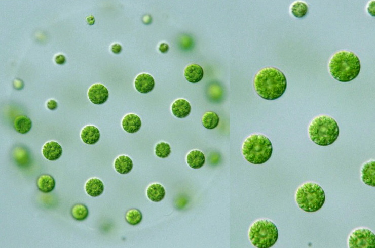
Pleodorina californica Shaw: Colonies of 128 cells, of which 48-64 cells (anterior 1/3~1/2 of the cells in a colony) small, 8-15 μm in diam., others large, 30-35 μm in diam.; colonies spherical in shape, 150-200 μm in diam.; cells with a single cup-shaped pyrenoid; (Illustrations of The Japanese Fresh-water Algae, 1977).
* Pleodorina japonica Nozaki: Colonies of 128 or 64 cells arranged at the perphery of a gelatinous matrix(up to 300 μm long); anterior 1/5~1/4 of the cells in a colony small, obligately somatic, nearly spherical (up to 16 μm in diam.; remaining cells reproductive, large (up to 30 μm in diam.); both with two equal-length flagella, a stigma and many contractile vacuoles; reproductive cells with many pyrenoids, somatic cells with a single pyrenoid (Photomicrographs of the Freshwater Algae, vol. 10, 1989).
Pleodorina indica (Iyengar) Nozaki: Colonies of 128 or 64 or 32 cells arranged at the perphery of a gelatinous matrix(up to 300 μm long); anterior 1/5~1/3 of the cells in a colony small, obligately somatic, nearly spherical (up to 16 μm in diam.; remaining cells gradually increase in cell size from anterior to posterior pole (up to 25 μm in diam.); gelatinous matrix forming individual sheaths (Photomicrographs of the Freshwater Algae, vol. 10, 1989).