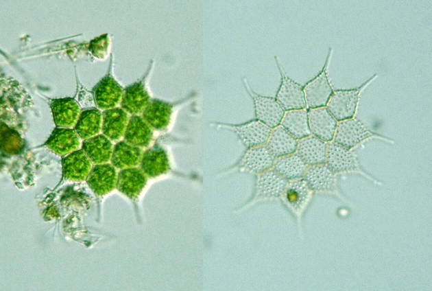
 Genus: Colonies of a fixed number of cells, flat, circular in shape; cell body polygonal in shape,
with horn-like projections; cell wall garanulated, wrinkled or notched;
chloroplasts plate-like or reticular; asexual reproduction by zoospore; sexual reproduction by isogametes
(Illustrations of The Japanese Fresh-water Algae, 1977).
Species: Marginal cells with a single, long horn-like process (Photomicrographs of the Freshwater Algae, vol. 8, 1988).
Var. echinulatum: Colonies without perforations; cell wall densely covered with smallspines (Photomicrographs of the Freshwater Algae, vol. 11, 1988).
Var. simplex: Colonies of 4, 8, 16, 32 cells, intercellular space absent, cells 20-30 μm long, 6-15 μm wide (Illustrations of The Japanese Fresh-water Algae, 1977).


