
Chlorophyceae:
Chlorococcales: Oocystaceae
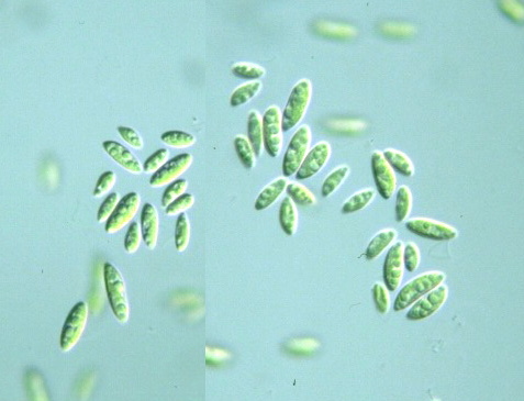
Monoraphidium lunare
Nygaard, Komárek, Kristiansen & Skulberg
 Genus: Cell body elongated spindle in shape, straight or curved, sharply pointed at both ends;
(Guide book to the "Photomicrographs of the Freshwater Algae", 1998).
Species: Cell body spindle-shaped, moderately curved, with blunt ends,
8-17 μm long, 1.5-3.5 μm wide;
a chloroplast parietal with a naked pyrenoid (invisible under photomicroscope);
cell division produces 4 or 8 "autospores"
(Photomicrographs of the Freshwater Algae, 1997).
|
Monoraphidium lunare, stock Sto-1, axenic culture with KCM and Hyponex (February 18 to March 23, 2002),
cell body 7.5-15 μm long, 4-5 μm wide,
x 640, Sayama Natural Park, Higashi-murayama city, Tokyo, Japan, December 2001 by Y. Tsukii
 50 μm
50 μm
 100 μm
 150 μm; x 400 :
 31 μm
31 μm
 63 μm
63 μm
 94 μm; x 640


Monoraphidium lunare,
stock Sto-1, axenic culture with KCM & Hyponex (January 25 to February 26, 2003),
cell body μm long, μm wide,
x 640, Sayama Natural Park, Higashi-murayama city, Tokyo, Japan, December 2001 by Y. Tsukii
 31 μm
31 μm
 63 μm
63 μm
 94 μm; x 640


Monoraphidium lunare,
stock Sto-01, axenic culture with KCM & Hyponex plus alpha (February 22 to April 5, 2003),
cell body μm long, μm wide,
x 640, Sayama Natural Park, Tokorozawa city, Saitama Pref., Japan, January 2003 by Y. Tsukii
 31 μm
31 μm
 63 μm
63 μm
 94 μm; x 640


Monoraphidium litorale Hindak:
Cells singular, fusiform, often attached to a subtrate or to mother cell wall remnant with one end by means of
a mucous target; 22-43 micron long, 2-6 micron wide; a chloroplast pariental, trough-shaped,
with a naked pyrenoid (without starch-envelope); cell division produces 4, 8 or 16 autospores (daughter cells)
releasing by crosswise rupture of mother cell wall
(Phtomicrog. Freshw. Alg. 16: 56, 1996)
Please click on images for viewing enlarged.
Copyright
Protist Information Server
 Genus: Cell body elongated spindle in shape, straight or curved, sharply pointed at both ends;
(Guide book to the "Photomicrographs of the Freshwater Algae", 1998).


