|
var. dianae (?) : L/W less than 10 |

v. dianae
190 x 24 μm
170 x 21 μm
L/W=8.1-10.0
Igashira park
Mooka
Tochigi, 2004 |

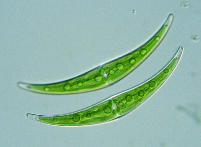
v. dianae
160 x 17 μm
L/W=9.4
Kuroiso Park
Kuroiso
Tochigi, 2003 |

v. dianae
155 x 17 μm
L/W=9.1
Lake Shirakaba
Tateshina & Chino
Nagano, 2005 |

v. dianae
or C. tumidulum ?
130 x 16 μm
L/W=8.4
Saitama, 2001 |

v. dianae
190 x 24 μm
L/W=7.9
Iruma river
Kawagoe
Saitama, 2004 |

v. dianae
152 x 20 μm
L/W=7.6
Koigakubo marsh
Tessei-cho
Okayama, 2004 |

v. dianae ?
150 x 20 μm
L/W=7.5
Imori pond
Myoko highland
Niigata, 2004 |

v. dianae ?
145, 170, 178 μm
L/W=7.3
Minuma-tanbo
Urawa
Saitama, 2001 |

v. dianae
160 x 17 μm
L/W=9.4
Lake Tanuki
Fujinomiya
Shizuoka, 2006
 |

v. dianae
148 x 16 μm
L/W=9.3
near Omine mt.
Minakami
Gunma, 2006
|