 Species: Cell body broad ellipsoidal or nearly spherical, 18-35 μm in diam.;
papilla broad with flattened apex; chloroplast cup-shaped; a pyrenoid horseshoe-shaped, located
beneath equatorial line or slightly posterior; stigma longitudinally elongated, thin,
located at equator or slightly anterior; nucleus at anterior half of the cell body.
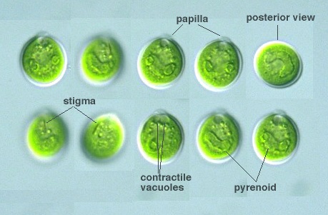
Species: Cell body broad ellipsoidal or nearly spherical, 18-35 μm in diam.;
papilla broad with flattened apex; chloroplast cup-shaped; a pyrenoid horseshoe-shaped, located
beneath equatorial line or slightly posterior; stigma longitudinally elongated, thin,
located at equator or slightly anterior; nucleus at anterior half of the cell body. [ var. globulifera (Korschikoff) Korschikoff 1938]: Papilla narrower and more protruded; cellwall thick; with several pyrenoids irregular in shape. [ var. perforata Vlk 1940]: Pyrenoid ring-form. [ var. longirubra Ettl 1976]: Stigma large, elongated, stick-like in shape, longitudinally located at anterior half of the cell; papilla more broad and short; a pyrenoid elongated, curved as half-ring (Süßwasserflora von Mitteleuropa 9, Chlorophyta I, 1983). |


An algal parasite, Rhyzophydium sp. (Chytridiales, Chytridiomycota, Eumycota) attached Chlamydomonas monadina,
C. monadina cell body 17 μm long, 16 μm wide,
Rhyzophydium cell body spherical 2 μm in diam.,
x 640, Akigase Park, Saitama city, Saitama Pref., Japan, March 2002 by Y. Tsukii
![]() 31 μm
31 μm
![]() 63 μm
63 μm
![]() 94 μm; x 640
94 μm; x 640


