 Species:
Cell body spherical in shape, 6-8 μm in diam.; papilla absent;
chloroplast cup-shaped, with a large anterior concavity; a single pyrenoid located at posterior half of the cell body;
stigma circular in shape, located at equator; nucleus at anterior
(Süßwasserflora von Mitteleuropa 9, Chlorophyta I, 1983).
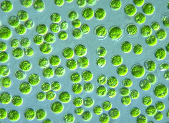
Species:
Cell body spherical in shape, 6-8 μm in diam.; papilla absent;
chloroplast cup-shaped, with a large anterior concavity; a single pyrenoid located at posterior half of the cell body;
stigma circular in shape, located at equator; nucleus at anterior
(Süßwasserflora von Mitteleuropa 9, Chlorophyta I, 1983).
|


