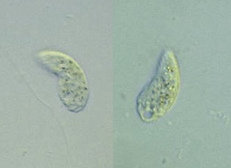
 Genus: Body reniform; dorsoventrally flattened; right body edge convex, left concave;
somatic groove originates on the dorsal surface, travels around the left side to the entrace of the
vestibulum on the ventral surface (Carey, 1992).
Oral cavity not tubular (Illustrated Guide, 1985).
Genus: Body reniform; dorsoventrally flattened; right body edge convex, left concave;
somatic groove originates on the dorsal surface, travels around the left side to the entrace of the
vestibulum on the ventral surface (Carey, 1992).
Oral cavity not tubular (Illustrated Guide, 1985).
Species: Cell body 50 μm long (Kahl, 1930). |


C. maupasi Enriques, 1908: 35-80 μm long;
20-50 μm wide;
resting cysts 21-28 μm diam.
(Foissner, Blatterer, Berger & Kohmann, 1991).
C. aspera Kahl, 1926: 30-40 μm long;
reproductive cysts with 4 tomites
(Foissner, Blatterer, Berger & Kohmann, 1991).
C. steinii Maupas, 1883: Renifrom; cytostomial cleft 1/3 of the body length from the
anterior end; 15-42 μm long;
smatic ciliation in 10 rows; 2 caudal cilia; vestibulum equipped with a beard of
'pseudomembranelles' arising from posterior margin; a macronucleus ovoid (Carey, 1992).
20-40 μm long;
15-30 μm wide; but size is quite variable
(Foissner, Blatterer, Berger & Kohmann, 1991).
15-40 μm; two long posterior cilia
(How to know the protozoa, 1979).
C. ecaudata (Liebmann, 1936): 20-40 μm long
(Foissner, Blatterer, Berger & Kohmann, 1991).
C. edaphoni Foissner, 1980: 25-40 μm long;
12-16 μm wide
(Foissner, Blatterer, Berger & Kohmann, 1991).
C. ellioti Bradbury & Outka, 1967: 15-35 μm long;
7-18 μm wide
(Foissner, Blatterer, Berger & Kohmann, 1991).