
Gamophyceae:
Zygnematales: Desmidiaceae or Closteriaceae
Closterium kuetzingii
Brébisson var. kuetzingii
 Species: Cell body quickly attenuated toward both ends (cf. C. ralfsii),
each extended part shorter than spindle-shaped part at middle (cf. C. setaceum),
both termini slighlty curved, rounded, 270-690 μm long, 14-27 μm wide ( L/W= 18-20);
cell wall transparent or brownish with thin lines (8-10 lines/10 μm wide);
chloroplasts composed of 5 lamellae; 4-6 pyrenoids in each half-cell
(Illustrations of The Japanese Fresh-water Algae, 1977).
270-512 μm long, 13-22 μm wide ( L/W= 18-30); 8-11 lines/10 μm wide
(Photomicrographs of the Fresh-water Algae, vol. 4, 1985).
|
Similar species -->>
C. ralfsii;
C. setaceum;
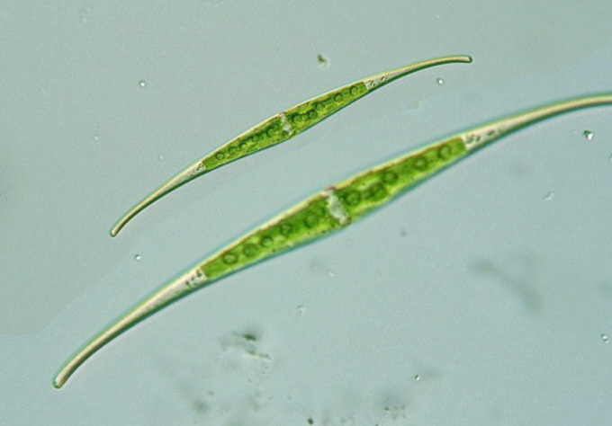
Closterium kuetzingii Brébisson var. kuetzingii,
cell body 200 μm long, 15 μm wide,L/W=13.3,
cell wall brownish in color, finely striated (see 59.jpg or 60.jpg),
x 640, Koigakubo marsh, Tessei-cho, Okayama Pref., Japan, November 22, 2004 by Y. Tsukii
 50 μm
50 μm
 100 μm
 150 μm; x 400 :
 31 μm
31 μm
 63 μm
63 μm
 94 μm; x 640


Cell body 205 μm long, 14 μm wide, L/W=14.6, x 400, x 640


Closterium setaceum Ehrenberg var. setaceum f. setaceum:
Cell body small in size, elongate, L/W= 30-42,
252-528 μm long,
6-16 μm wide;
both termini slightly curved, spindle-shaped at middle; cell wall transparent or brownish
with thin lines (8-13 lines/10 μm wide);
2-3 pyrenoids in each half-cell (Photomicrographs of the Fresh-water Algae, vol. 6, 1997).
* Closterium kuetzingii BrébissonBrébisson var.
kuetzingii: Cell body moderate in size, elongate, L/W= 18-30,
270-512 μm long,
13-22 μm wide;
both termini slightly curved, spindle-shaped at middle; cell wall transparent or brownish
with thin lines (8-11 lines/10 μm wide);
chloroplasts composed of 5 lamellae; 4-6 pyrenoids in each half-cell
(Photomicrographs of the Fresh-water Algae, vol. 4, 1985).
Please click on images for viewing enlarged.
Copyright
Protist Information Server
 Species: Cell body quickly attenuated toward both ends (cf. C. ralfsii),
each extended part shorter than spindle-shaped part at middle (cf. C. setaceum),
both termini slighlty curved, rounded, 270-690 μm long, 14-27 μm wide ( L/W= 18-20);
cell wall transparent or brownish with thin lines (8-10 lines/10 μm wide);
chloroplasts composed of 5 lamellae; 4-6 pyrenoids in each half-cell
(Illustrations of The Japanese Fresh-water Algae, 1977).


